How 3D Pathomorphology Enhances Anatomy Education
Pathology education has traditionally relied on microscopic examination of tissue samples and two-dimensional illustrations in textbooks. While microscopy remains essential for understanding cellular-level changes, students also need to understand how diseases manifest at the organ and system level. Three-dimensional pathomorphology models address this gap by providing interactive, manipulable representations of diseased organs.
From 2D Slides to 3D Understanding
When a student looks at a histology slide showing hepatic fibrosis, they see tissue-level changes. But understanding how that fibrosis affects the overall architecture of the liver, how it alters blood flow through the portal system, and how it leads to the clinical signs of cirrhosis requires a macroscopic perspective. 3D pathomorphology models bridge this gap by showing the organ in its complete form, with pathological changes visible in three dimensions.
Students can rotate the model, zoom in on specific regions, and compare healthy and diseased states side by side. This interactive exploration builds the spatial understanding that textbook diagrams cannot fully convey.
Coverage Across Specialties
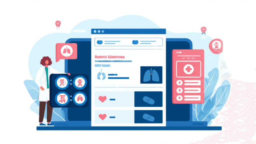
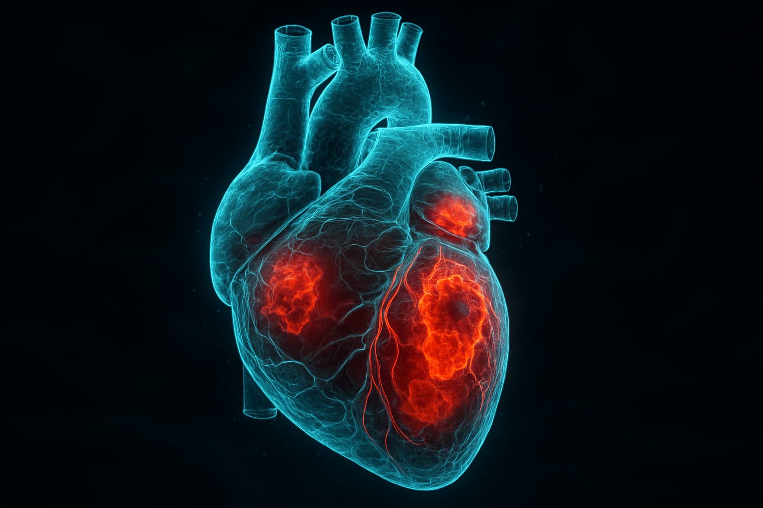
A comprehensive pathomorphology library covers target organs across medical specialties. Heart models demonstrate cardiomyopathies, valvular disease, and coronary artery pathology. Lung models show emphysema, pneumonia, and tumor growth patterns. Kidney models illustrate glomerulonephritis, polycystic disease, and hydronephrosis. Each model represents conditions that students will encounter in clinical practice.
When a platform provides models covering multiple organ systems, students can explore connections between specialties. They can see how systemic diseases like diabetes affect the kidneys, eyes, and peripheral nerves simultaneously, building the integrative thinking that characterizes skilled clinicians.
Integration with Clinical Cases
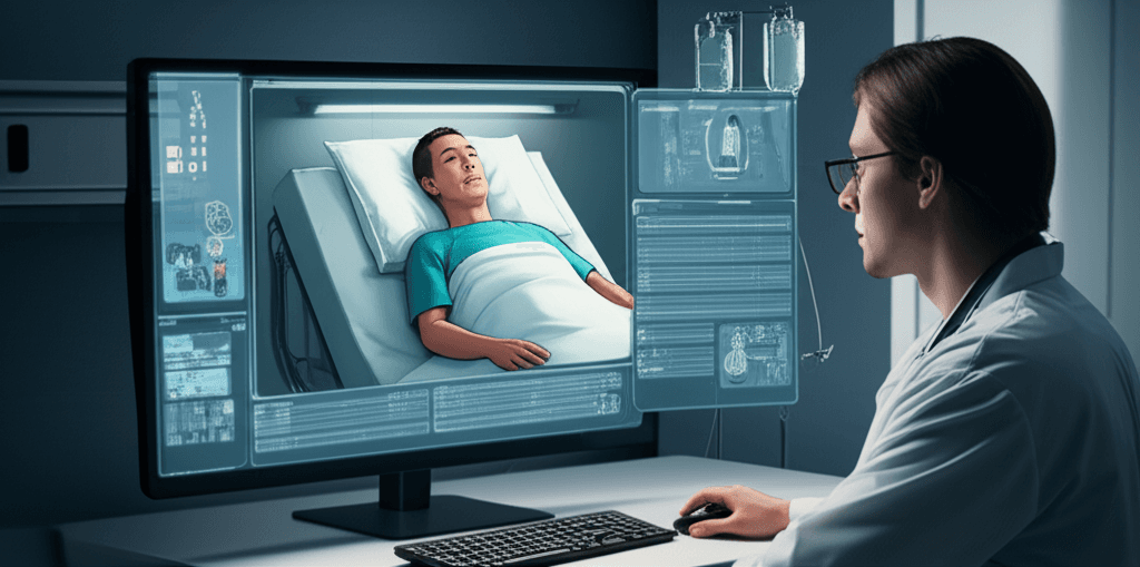
3D pathomorphology becomes most powerful when integrated with clinical case scenarios. When a student is working through a virtual patient case involving chest pain, they can step into the pathomorphology mode to examine a 3D model of the heart, visualize coronary artery stenosis, and understand the anatomical basis of the symptoms they are evaluating. This connection between clinical presentation and underlying pathology reinforces learning in both directions.
Advantages Over Traditional Methods
Cadaver dissection provides a three-dimensional perspective but is limited by availability, cost, and the fact that cadavers typically show age-related changes rather than specific pathological conditions. Digital 3D models can represent any condition at any stage of progression, are infinitely reusable, and can be accessed at any time from any location with a computer or VR headset.
The VR application of pathomorphology is particularly compelling. When students can hold a virtual organ in their hands, rotate it naturally, and examine it from any angle, the learning experience approaches the spatial richness of physical examination while offering the pathological specificity that cadavers lack.
Practical Implementation for Educators
For medical schools considering 3D pathomorphology tools, the key evaluation criteria are the accuracy of anatomical models, the range of pathological conditions represented, the quality of the rendering and interaction, and the ease of integration into existing curricula. The platform should work across devices (desktop, web, VR) so that students can access models both in structured lab sessions and during independent study.
The most effective approach uses 3D pathomorphology as a complement to, not a replacement for, traditional pathology teaching. Students who study histology slides in the lab and then explore 3D organ models of the same conditions develop both microscopic and macroscopic understanding of disease processes.