The Role of 3D Anatomy Models in Modern Medical Education
Anatomy is the bedrock of medical education. Every clinical specialty depends on a thorough understanding of human body structure. For centuries, cadaver dissection has been the primary method for teaching anatomy, and it remains valuable. However, cadaver programs face practical limitations that 3D digital anatomy models are uniquely positioned to address.
Limitations of Traditional Anatomy Teaching
Cadaver dissection programs are expensive to operate, requiring dedicated facilities with specialized ventilation, preservation chemicals, and trained prosectors. The supply of cadavers varies by region, and some institutions face chronic shortages. Each cadaver can be dissected once, and the process is irreversible. Students who miss a dissection session cannot recreate the experience.
Two-dimensional textbook illustrations, while high quality, lack the spatial relationships that three-dimensional structures naturally possess. Students studying the brachial plexus in a textbook see a flat diagram; the actual nerve network occupies three-dimensional space in relation to muscles, bones, and blood vessels. This spatial relationship is critical for surgical planning and clinical examination.
What 3D Anatomy Models Offer
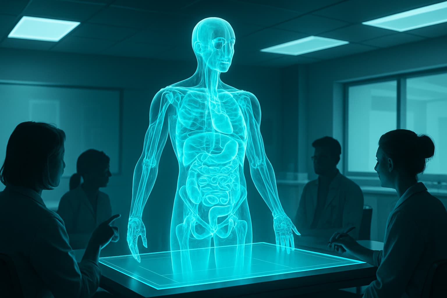
Interactive 3D anatomy applications allow students to explore a complete human body model at their own pace. They can isolate individual structures (showing only the cardiovascular system, for example), rotate the view to any angle, zoom from whole-body overview to microscopic detail, and toggle between different systems to understand their spatial relationships.
The ability to show and hide structures is particularly powerful. A student studying the relationship between the thoracic aorta and the esophagus can hide overlying structures to reveal the relevant anatomy, then restore them to understand context. This layer-by-layer exploration is impossible with a physical cadaver, where removing one structure permanently alters the remaining anatomy.
Accessibility and Repetition
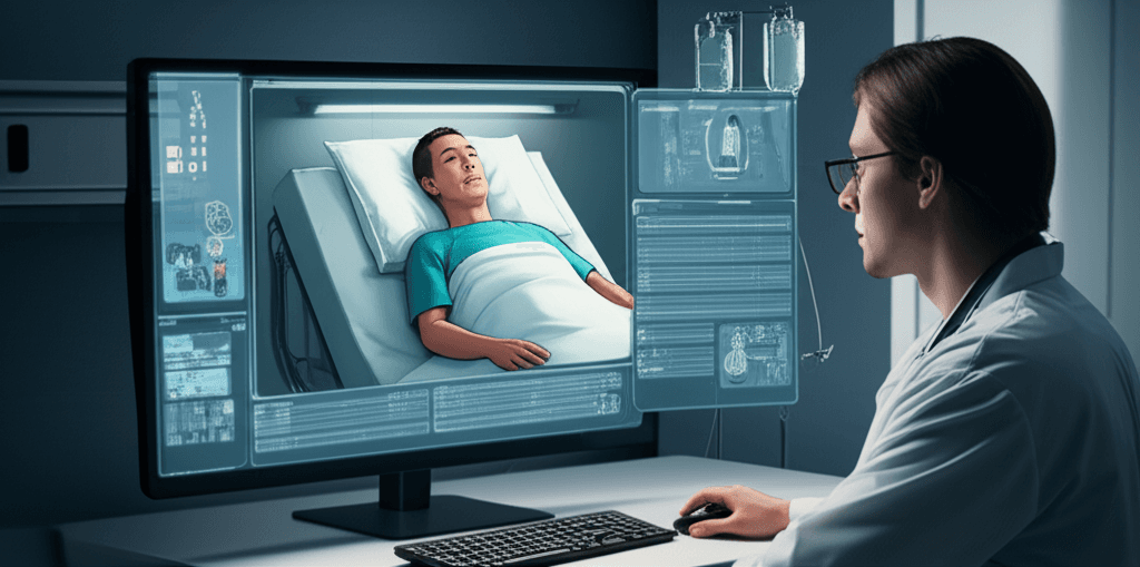
Digital anatomy models are available anytime, anywhere. A student can study cardiac anatomy on a laptop at home, on a tablet in the library, or in a VR headset in the simulation lab. This accessibility dramatically increases the total hours of anatomy study available to each student compared to scheduled cadaver lab sessions.
Repetition is equally important. A student preparing for a surgery rotation can review the anatomy of the inguinal canal as many times as needed. In a cadaver lab, that region may have already been dissected and is no longer available for further study. Digital models never degrade, are never damaged by inexpert dissection, and are always available in their original state.
Clinical Correlation: Anatomy in Context
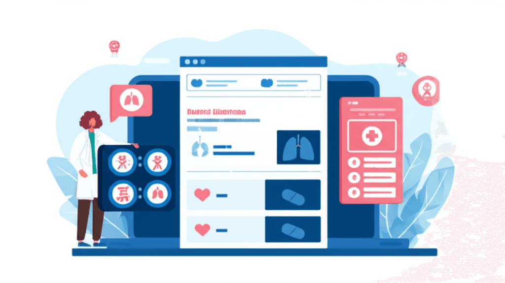
The most effective 3D anatomy tools integrate with clinical content. When a student is studying the anatomy of the knee, the platform can link to relevant clinical cases (meniscal tears, ACL injuries, osteoarthritis), relevant imaging (MRI sequences), and relevant procedures (arthroscopy steps). This clinical correlation makes anatomy study immediately relevant to medical practice rather than an abstract memorization exercise.
This integration is where comprehensive medical simulation platforms add value beyond standalone anatomy applications. When the same platform offers both 3D anatomy and virtual patient cases, students can move seamlessly between anatomical study and clinical application.
Complementing, Not Replacing, Cadaver Dissection
The evidence-based consensus in medical education is that 3D digital anatomy complements rather than replaces cadaver dissection. Cadavers provide tactile experience, tissue texture, natural anatomical variation, and the emotional component of working with human remains that digital tools cannot replicate.
The most effective anatomy programs use both: digital 3D models for initial learning, spatial understanding, and unlimited review, combined with cadaver dissection for hands-on experience and the irreplaceable reality of human tissue. Students who arrive at the cadaver lab having already studied the anatomy in 3D use their limited dissection time more effectively.